How TMS Works on the Brain Part 11:The Role of Personalized Targeting in TMS
One of the most transformative developments in transcranial magnetic stimulation (TMS) is the rise of personalized targeting. Traditional TMS protocols often use standardized scalp measurements to locate treatment sites—such as positioning the coil a set distance forward from the motor cortex to reach the dorsolateral prefrontal cortex (DLPFC). While effective for many, this approach assumes all brains are wired the same, which we now know isn’t true.
Thanks to advances in brain imaging and neuroscience, it’s become increasingly clear that psychiatric disorders like depression, anxiety, and OCD stem from dysfunctional brain networks that vary from person to person. Personalized TMS seeks to tailor treatment based on each individual’s unique brain structure and functional connectivity, aiming for better accuracy—and better outcomes.
Why Standard Targeting Falls Short
Standardized TMS relies on surface landmarks and population averages, which can miss the most therapeutically relevant brain regions for a given patient. Anatomical differences, previous injuries, or atypical wiring can shift where key circuits actually reside. Even among patients with the same diagnosis, the underlying network dysfunction can differ dramatically.
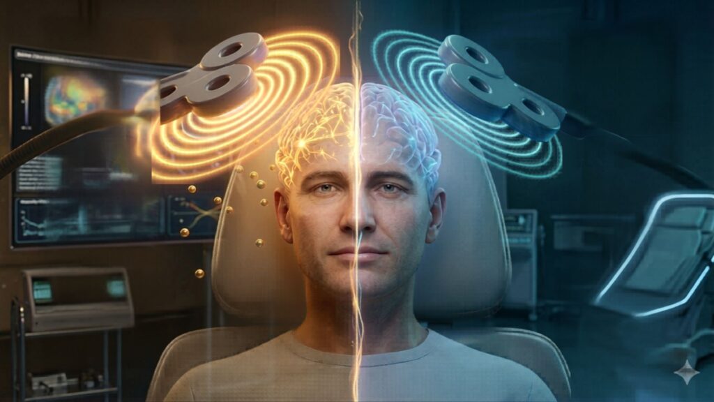
For example, one person with depression may have hypoactivity in the left DLPFC, while another may show abnormal connectivity between the prefrontal cortex and deeper limbic structures. Using the same treatment site for both might not yield equally strong results.
How Personalized Targeting Works
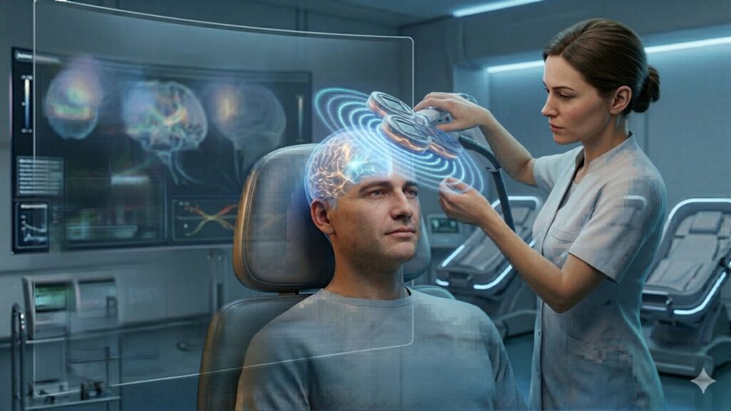
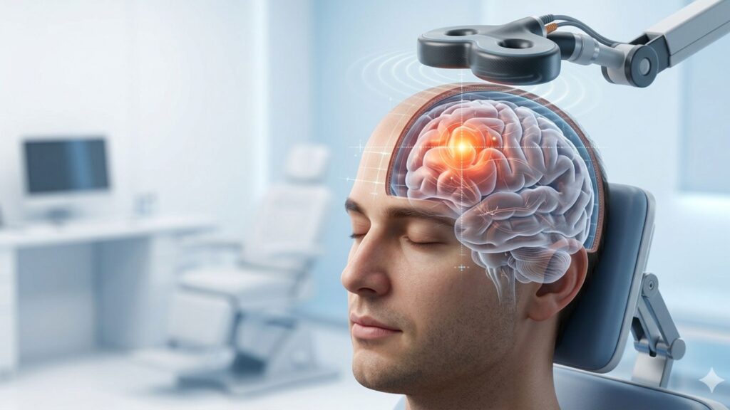
Personalized TMS begins with a detailed brain map. Clinicians may use structural MRI, functional MRI (fMRI), or quantitative EEG (qEEG) to understand a patient’s individual brain anatomy and activity. These tools help identify which areas are underactive, overactive, or improperly connected—and guide where to stimulate for optimal results.
In connectivity-based targeting, for example, researchers look for brain regions with strong functional connections to key emotional processing hubs like the subgenual anterior cingulate cortex (sgACC). Stimulating areas with stronger connectivity to the sgACC has been shown to improve outcomes in treatment-resistant depression.
qEEG-guided targeting is another promising approach. By analyzing patterns of brainwave activity, clinicians can pinpoint dysregulated regions, assess hemispheric imbalances, and determine whether high- or low-frequency stimulation is most appropriate. This method is especially valuable for conditions like ADHD, PTSD, or autism spectrum disorder, where individual brain profiles vary widely.
The Evidence Behind Personalized TMS
Several studies have now confirmed that tailoring TMS based on functional connectivity or individual biomarkers can lead to better clinical outcomes. A landmark study by Cash et al. (2021) found that patients whose treatment sites were chosen based on their brain’s connectivity to the sgACC experienced greater reductions in depressive symptoms than those treated at standard locations.
Similarly, personalized approaches using qEEG have shown potential in reducing symptom severity across a range of disorders. Early research in autism, for instance, suggests that custom-mapped stimulation may enhance social cognition and executive function more effectively than fixed-site protocols. Studies using qEEG have found that children and adults with autism often show atypical patterns of brainwave activity—such as excess theta or reduced alpha rhythms—particularly in regions associated with attention, language, and social interaction. Targeting these specific dysregulated areas with TMS may help normalize brainwave activity and improve behavioral outcomes.
Practical Applications and Tools
Clinics that offer personalized TMS typically integrate neuronavigation systems into their workflow. These systems overlay a patient’s MRI or EEG data onto a 3D model of the brain, allowing the clinician to position the coil with millimeter precision. Some platforms also provide electric field modeling, which predicts how stimulation will spread through brain tissue—further refining target selection.
Although more time-consuming and resource-intensive, personalized targeting is increasingly seen as the gold standard for complex or treatment-resistant cases. As these tools become more accessible and cost-effective, it’s likely they’ll play a larger role in standard clinical practice.
Challenges and the Path Forward
Despite its promise, personalized TMS is not without challenges. High-quality imaging requires specialized equipment and trained personnel, which may not be available in every clinic. Insurance coverage for imaging-guided protocols also remains inconsistent. Furthermore, ongoing research is needed to refine targeting algorithms and determine which biomarkers are most predictive of treatment response.
Still, the field is advancing rapidly. Machine learning and artificial intelligence are being used to analyze vast datasets and predict optimal targets based on a combination of symptoms, brain structure, and connectivity. In the near future, personalized TMS could become faster, more automated, and more widely available.
Conclusion
Personalized targeting represents the next frontier in TMS. By recognizing the uniqueness of every brain and tailoring treatment accordingly, clinicians can achieve better outcomes with fewer sessions and less trial and error. As technology and research continue to evolve, personalized TMS is poised to become the norm—bringing precision medicine to the forefront of mental health care.
References
- Cash, R. F. H., Cocchi, L., Lv, J., Wu, Y., Fitzgerald, P. B., & Zalesky, A. (2021). Personalized connectivity-guided DLPFC-TMS for depression: Advancing computational feasibility, precision and reproducibility. Human brain mapping, 42(13), 4155–4172. https://doi.org/10.1002/hbm.25330
- Fox, M. D., Buckner, R. L., White, M. P., Greicius, M. D., & Pascual-Leone, A. (2012). Efficacy of transcranial magnetic stimulation targets for depression is related to intrinsic functional connectivity with the subgenual cingulate. Biological Psychiatry, 72(7), 595–603. https://doi.org/10.1016/j.biopsych.2012.04.028
- van Bueren, N. E. R., Reed, T. L., Nguyen, V., Sheffield, J. G., van der Ven, S. H. G., Osborne, M. A., Kroesbergen, E. H., & Cohen Kadosh, R. (2021). Personalized brain stimulation for effective neurointervention across participants. PLoS computational biology, 17(9), e1008886. https://doi.org/10.1371/journal.pcbi.1008886