How TMS Works on the Brain Part 5:Targeting Specific Brain Regions with TMS
One of the most powerful aspects of transcranial magnetic stimulation (TMS) is its ability to precisely target specific regions of the brain. Because different brain areas are responsible for various cognitive, emotional, and motor functions, the therapeutic outcome of TMS greatly depends on where the stimulation is delivered. This flexibility allows clinicians to treat a wide range of disorders, from depression and anxiety to chronic pain and neurological impairments.
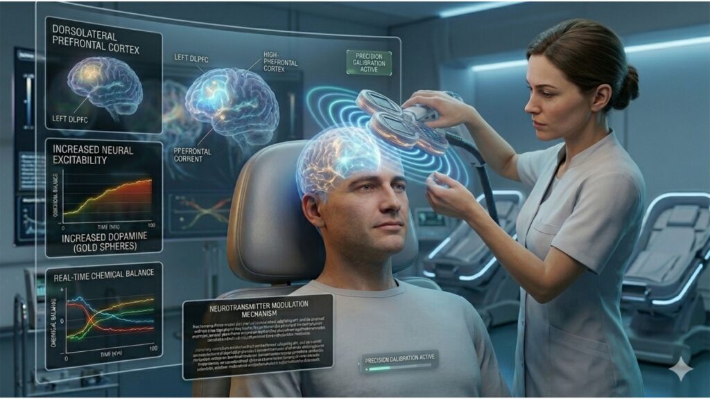
The most commonly targeted region in clinical practice is the dorsolateral prefrontal cortex (DLPFC). This area, particularly on the left side, plays a key role in emotional regulation, decision-making, and motivation. In individuals with depression, activity in the left DLPFC is often reduced. High-frequency TMS applied to this area helps restore normal activity, which is associated with improved mood, concentration, and executive function. For this reason, left DLPFC stimulation remains the gold standard for treating major depressive disorder (MDD).
On the other hand, the right DLPFC is frequently targeted in anxiety disorders. Low-frequency stimulation of this region helps reduce hyperactivity that is often associated with worry, fear, and panic. This inhibitory approach has demonstrated effectiveness in conditions such as generalized anxiety disorder (GAD), panic disorder, and post-traumatic stress disorder (PTSD). By calming overactive circuits, patients often experience reduced emotional reactivity and better control over anxious thoughts.
Beyond mood and anxiety disorders, TMS is increasingly used to modulate brain regions tied to other psychiatric and neurological conditions. For instance, the supplementary motor area (SMA) is a key target in the treatment of obsessive-compulsive disorder (OCD). The SMA is involved in motor planning and behavioral inhibition. In individuals with OCD, this area is often overactive, contributing to the compulsive drive to perform rituals and routines. Modulating the SMA through TMS has shown promise in reducing compulsive behaviors and improving symptom control, particularly when paired with cognitive-behavioral therapy (CBT).
In patients experiencing auditory hallucinations associated with schizophrenia, clinicians often direct low-frequency TMS to the left temporoparietal cortex, a region involved in auditory processing and language comprehension. Research suggests that overstimulation in this area may contribute to the perception of internally generated voices. TMS can dampen this overactivity, reducing the intensity and frequency of hallucinations in treatment-resistant cases.
The application of TMS in chronic pain and motor recovery, particularly after a stroke, also continues to grow. One of the primary stimulation sites for these conditions is the primary motor cortex (M1). High-frequency stimulation over M1 is believed to reduce pain perception by modulating both local and descending pain pathways. For stroke patients, stimulating motor regions of the affected hemisphere may help recruit surrounding neurons, supporting neuroplasticity and aiding in motor rehabilitation, especially when paired with physical therapy or occupational therapy interventions.
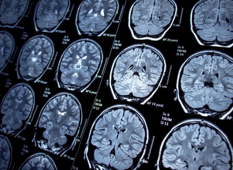
As our understanding of brain connectivity evolves, so does our ability to optimize TMS treatment using advanced imaging techniques. Tools such as functional magnetic resonance imaging (fMRI), electroencephalography (EEG), and diffusion tensor imaging (DTI) help identify abnormalities in structure or function. These technologies allow clinicians to tailor treatment protocols based on the unique neuroanatomy and network dysfunctions of each patient.
To support this level of personalization, many clinics use neuronavigation systems—a technique that overlays a patient’s brain map with real-time data to ensure the TMS coil is positioned with millimeter precision. Neuronavigation has become a standard for complex or treatment-resistant cases, improving both consistency and effectiveness. For example, rather than relying on scalp measurements to locate the DLPFC, a clinician can directly stimulate the precise coordinates of hypoactive tissue based on a patient’s MRI scan. This method is especially useful when standard protocols have failed or when anatomical variations are present.
In recent years, interest has expanded around deep TMS (dTMS), which uses H-coils designed to penetrate deeper brain structures than traditional figure-eight coils. These coils allow for broader stimulation zones, reaching areas such as the anterior cingulate cortex (ACC) and insula, both of which are involved in mood regulation, pain perception, and emotional processing. The ACC, in particular, is a critical node in the brain’s salience network, and dysfunction here has been linked to treatment-resistant depression. Deep TMS targeting this region has shown promise not only for depression but also for smoking cessation, OCD, and bipolar depression.
Researchers are also investigating the use of deep TMS in treating addiction, where disruptions in reward pathways involving the prefrontal cortex and limbic system play a central role. Early trials suggest that repeated stimulation of these areas may reduce cravings and promote behavioral regulation, offering a novel approach for individuals with substance use disorders who have not responded to conventional treatment.
As TMS technology continues to evolve, so too does its list of potential applications. New protocols are being tested for autism spectrum disorder, ADHD, and even Alzheimer’s disease. In each case, precise targeting of affected neural circuits is central to the approach. The future of TMS lies not only in expanding the list of treatable conditions but also in enhancing our ability to personalize and refine stimulation strategies.
Ultimately, the ability to target specific brain regions is what gives TMS its wide-ranging therapeutic potential. Whether stimulating the DLPFC to lift mood, calming the SMA to reduce compulsions, or reaching into deeper limbic structures to stabilize emotional regulation, the impact of TMS is deeply tied to its anatomical precision. Combined with modern imaging and navigation tools, this capability positions TMS as one of the most adaptable and promising treatments in neuroscience today.
References
- Fox, M. D., Buckner, R. L., White, M. P., Greicius, M. D., & Pascual-Leone, A. (2012). Efficacy of transcranial magnetic stimulation targets for depression is related to intrinsic functional connectivity with the subgenual cingulate. Biological Psychiatry, 72(7), 595–603. https://doi.org/10.1016/j.biopsych.2012.04.028
- Lefaucheur, J. P., Aleman, A., Baeken, C., et al. (2020). Evidence-based guidelines on the therapeutic use of repetitive transcranial magnetic stimulation (rTMS). Clinical Neurophysiology, 131(2), 474–528. https://doi.org/10.1016/j.clinph.2019.11.002
- Slotema, C. W., Blom, J. D., Hoek, H. W., & Sommer, I. E. (2014). Should we expand the toolbox of psychiatric treatment methods to include rTMS? A meta-analysis of the efficacy of rTMS in psychiatric disorders. Journal of Clinical Psychiatry, 75(11), 1213–1222. https://pubmed.ncbi.nlm.nih.gov/20361902/
- Cash, R. F. H., Cocchi, L., Lv, J., Wu, Y., Fitzgerald, P. B., & Zalesky, A. (2021). Personalized connectivity-guided DLPFC-TMS for depression: Advancing computational feasibility, precision and reproducibility. Human brain mapping, 42(13), 4155–4172. https://doi.org/10.1002/hbm.25330